Histology Basics the microscopic study of tissues and organs through sectioning, staining examining
What you will learn
types of tissues
cardiovascular histology
respiratory histology
endocrine histology
reproductive histology
urinary histology
lymphoid histology
gastrointestinal histology
sensory histology
Description
Histology,[help 1] also known as microscopic anatomy or microanatomy, is the branch of biology that studies the microscopic anatomy of biological tissues Histology is the microscopic counterpart to gross anatomy, which looks at larger structures visible without a microscope.[5][6] Although one may divide microscopic anatomy into organology, the study of organs, histology, the study of tissues, and cytology, the study of cells, modern usage places all of these topics under the field of histology. In medicine, histopathology is the branch of histology that includes the microscopic identification and study of diseased tissue.
There are four basic types of animal tissues: muscle tissue, nervous tissue, connective tissue, and epithelial tissues
All animal tissues are considered to be subtypes of these four principal tissue types (for example, blood is classified as connective tissue, since the blood cells are suspended in an extracellular matrix, the plasma)
- Epithelium
- Simple epithelium
- Simple squamous epithelium
- Simple cuboidal epithelium
- Simple columnar epithelium
- Pseudostratified columnar epithelium
- Stratified epithelium
- Stratified squamous epithelium
- Stratified cuboidal epithelium
- Stratified columnar epithelium
- Transitional epithelium
- Multicellular glands
- Simple epithelium
- Muscle tissue
- Smooth muscle
- Skeletal muscle
- Cardiac muscle
- Connective tissue
- General connective tissue
- Loose connective tissue
- Dense connective tissue
- Special connective tissue
- Cartilage
- Bone
- Hemopoietic
- Blood
- Lymph
- General connective tissue
- Nervous tissue
- Central nervous system
- Peripheral nervous system
- Special receptors
Four basic types of human tissue can be stained and viewed using various histological techniques. Epithelium, connective tissue, muscle tissue, and nervous tissue have commonalities but look very distinct structurally after staining. Each stain exists to highlight an important feature or component within a tissue type. For example, one of the most common stains, Hematoxylin, is a basic dye that stains proteins a blue color, while Eosin stains proteins a pink color. These two stains are commonly used together to define intracellular organelles and proteins. Because of the variety of the proteins that exist, some stains were created to highlight a particular protein, which this review will discuss in the following sections. The benefit of using a special stain is that it can highlight the specific protein very well. However, because of its specificity, the other structures will not be seen. For this reason, multiple slides will often be created from a given specimen so that multiple stains can be performed to gather the full range of needed information.
Almost all tissue stains are performed on tissue that has been removed from the body. However, in rare instances, very specialize stains called vital stains can work on tissue remaining in the body. These stains are used for the identification of specific types of tissue and identification of abnormal tissue, so a subsequent biopsy can be more accurate in obtaining abnormal tissue
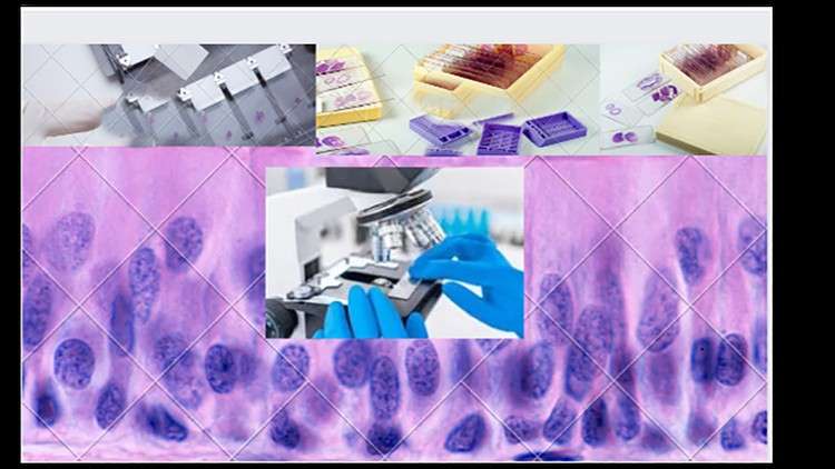
Tissue Preparation
Before specific staining can occur, tissue samples must undergo preparation through the following stages: Fixation, processing, embedding, sectioning, and sometimes antigen retrieval. In modern histology laboratories, most of these steps are automated.
.Fixation: Fixation uses chemicals to preserve the structure of the tissue in its natural form and protects it from degradation by irreversibly cross-linking proteins. Although several specialized fixatives are available, Neutral Buffered Formalin is a common choice for this step. The fixation step is vital to the rest of the histologic staining procedure because by retaining the chemical composition of the tissue, the sample is hardened and makes the sectioning phase easier. Paraffin-formalin is another effective fixative. Its benefit is that it is the fixative of choice for immunostaining; however, it requires preparation at the time of the fixation. Bouin is a fixative used for examining embryo and brain tissue because of its superior preservation of delicate nuclei and glycogen. Its downside is that it does not preserve kidney tissues well and also distorts mitochondrial structure.[1]
Dehydration: The addition of ethanol accomplishes the dehydration of a sample. It removed water from the sample and further hardens the tissue for eventual light microscopy. After ethanol is applied, and following the completion of tissue dehydration, xylene is used to remove the ethanol.[1]
Embedding: Embedding is the process of putting the sample into a paraffin wax or a plastic resin to enhance the process of extracting cellular structures. This step is to be performed with caution if the goal is to perform immunostaining because the paraffin wax will inhibit the penetration of antibodies, and lead to a false result.[1]
Sectioning: Sectioning involves mounting the specimen on a microtome and cutting it into sections. The preferred thickness is 4-5 micrometers so that it can be stained and put on a microscope slide for examination.[1]
Antigen Retrieval: This step is to retrieve antigens that could have been covered in the fixation and embedding stages. If the cross-linking of proteins conceals the antigen sites, there may not be as robust of an immunohistochemical response. Antigen retrieval is achieved through heating and proteolytic methods to break down the cross-links and reveal the epitopes and antigens that were previously covered.[1] Although this step carries the risk of denaturing both the fixative and the antigens themselves, a successful antigen retrieval method can lead to a much more effective immunostaining intensity.
Content
- Course Overview
- Unlock the intricate architecture of the human body at the cellular and tissue level through a captivating journey into microscopic anatomy.
- This course demystifies the art and science of histology, transforming complex visuals into easily understandable concepts.
- Gain a foundational understanding of how the microscopic structure of tissues dictates their function within various organ systems.
- Explore the fundamental building blocks of life and their arrangement, providing essential knowledge for a wide range of biological and medical disciplines.
- Develop a keen eye for distinguishing between different cellular morphologies and their characteristic tissue arrangements.
- Learn the principles behind preparing and visualizing tissue samples, bridging the gap between raw biological material and diagnostic insights.
- Master the ability to interpret microscopic images, a critical skill for accurate diagnosis and research.
- Requirements / Prerequisites
- A foundational understanding of basic biology and cell biology is beneficial but not strictly required.
- Curiosity and a willingness to engage with visual information are paramount.
- Access to a computer with internet connectivity for online lectures and resources.
- Basic familiarity with microscopy principles, though the course will reinforce these concepts.
- Skills Covered / Tools Used
- Microscopic Interpretation: Develop the ability to analyze and interpret histological slides, identifying key cellular and structural features.
- Staining Techniques: Understand the principles and applications of common histological stains and their role in visualizing cellular components.
- Tissue Identification: Learn to differentiate between major tissue types and their subtypes based on microscopic appearance.
- Spatial Reasoning: Enhance your ability to visualize three-dimensional tissue structures from two-dimensional microscopic sections.
- Digital Microscopy Tools: Familiarization with common digital microscopy platforms and image analysis software (as applicable to course materials).
- Comparative Anatomy (Microscopic): Learn to recognize subtle differences in tissue structure across different organ systems.
- Benefits / Outcomes
- Build a robust knowledge base essential for further studies in medicine, pathology, dentistry, nursing, and biomedical sciences.
- Significantly enhance your ability to understand and retain complex anatomical and physiological information.
- Gain a competitive edge in academic and professional settings requiring a strong grasp of microscopic anatomy.
- Empower yourself to critically evaluate scientific literature that relies on histological evidence.
- Develop a deeper appreciation for the elegance and efficiency of biological design at the microscopic level.
- Acquire a transferable skill set applicable to research, diagnostics, and clinical practice.
- PROS
- Accessibility: Designed for learners of all backgrounds, making a potentially daunting subject approachable.
- Practical Application: Focuses on skills directly applicable to a wide range of healthcare and research fields.
- Visual Learning Emphasis: Leverages visual aids and detailed explanations to solidify understanding.
- Comprehensive Scope: Covers key organ systems, providing a broad yet detailed overview.
- CONS
- Requires Dedicated Study: While made easy, mastery still necessitates consistent effort and review of microscopic details.